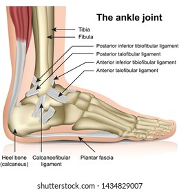
Tendon Diagram / A Patient S Guide To Foot Anatomy 2020 Orthonorcal Los Gatos Capitola Morgan Hill Watsonville Ca. Here you can see the tendons that extend down the top of your foot toward your toes, allowing you to curl your toes upward if need be. 2 ligaments (trapezoid& conoid ligaments) attach the clavicle coracoid process of scapula these tiny ligaments (w/ acominoclavicular joint) keep scapula attached to clavicle. Attaches the calf muscles to the calcaneus, most important muscles for running, jumping, walking etc. The foot diagram has a complex structure made up of bones, ligaments, muscles, and tendons.understanding the structure of the foot is best done by looking at a foot diagram where the anatomy has been labeled. The bones of the hip include the femur, the ilium, the ischium, and the pubis.
Related posts of foot tendons and ligaments diagram foot muscle blank diagram. Check out and click on the image to download it. The forearm is the part of your arm between the wrist and the elbow. Tendons, located at each end of a muscle, attach muscle to bone. One tendons inserts onto the forearm bone, the radius, and the second spreads out to join the fascia along the upper part of the forearm.
Knee Wikipedia from upload.wikimedia.org The current term that is recommended to describe this cohort of patients is 'tendinopathy'. Here you can see the tendons that extend down the top of your foot toward your toes, allowing you to curl your toes upward if need be. Pain in tendons between thumb and index finger. You may be able to treat forearm tendonitis with rest and. A tendon, also known as a sinew, is a fibrous tissue that helps to facilitate this movement. The pubis, ischium, and ilium together constitute the pelvis while the thigh bone is the femur. If you would like to learn all the parts of the foot structure, you have come to the right place. The achilles tendon is the largest.
Also allows the action of raising up onto toes.
You may be able to treat forearm tendonitis with rest and. Tendons that make this possible include: Related posts of shoulder muscles and tendons diagram neck muscle anatomy mri. Tendons play an important role in the movement by transmitting the contraction force produced by the muscles to the bone they hold, and their contribution to stability to the joints is extremely important. The tendon runs down the back of your lower leg from the back of the knee to the heel. Tendons attach muscles to bones. Ligaments and tendons serve similar purposes, but in different ways. They propose there are 3 stages to this continuum. Here you can see the tendons that extend down the top of your foot toward your toes, allowing you to curl your toes upward if need be. The foot diagram has a complex structure made up of bones, ligaments, muscles, and tendons.understanding the structure of the foot is best done by looking at a foot diagram where the anatomy has been labeled. Biceps tendons the biceps muscle has two tendons at the shoulder, called the long head and short head. Tendons are found throughout the body, from the head and neck all the way down to the feet. The bones together make up the hip.
Here you can see the tendons that extend down the top of your foot toward your toes, allowing you to curl your toes upward if need be. The achilles tendon is also called the calcaneal tendon. Foot muscle blank diagram 10 photos of the foot muscle blank diagram blank muscle diagram to label, blank muscle diagram worksheet, digestive system blank diagram, leg muscle diagram, muscular system blank diagram, respiratory system blank diagram, skeletal system blank diagram, foot, blank muscle diagram to. Your biceps tendons attach the biceps muscle to bones in the shoulder and in the elbow. The following diagram below is the human body muscle diagram.
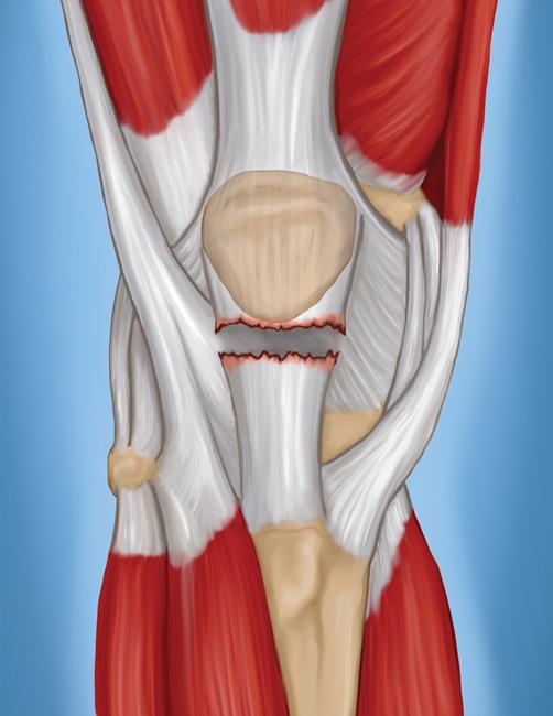
Ligament Hd Stock Images Shutterstock from image.shutterstock.com The muscle belly then crosses the entire upper arm and separates into two tendons. The forearm is the part of your arm between the wrist and the elbow. Check out and click on the image to download it. The fascicle contains the basic fibril of the ligament or tendon, and the fibroblasts, which are the biological cells that produce the ligament or tendon. Attaches the calf muscles to the calcaneus, most important muscles for running, jumping, walking etc. The patellar tendon connects the apex of the patella to the tibial tuberosity, and improves the way the quadriceps muscle pulls on the tibia. If you tear the biceps tendon at the shoulder, you may lose some strength in your arm and have pain when you forcefully turn your arm from palm down to palm up. Ligaments and tendons serve similar purposes, but in different ways.
The patella is a sesamoid bone that lies within the quadriceps tendon.
Tendons that make this possible include: One of the most important tendons in terms of mobility of the leg is the achilles tendon. The patella is a sesamoid bone that lies within the quadriceps tendon. Tendons are sometimes confused with ligaments. Foot muscle blank diagram 10 photos of the foot muscle blank diagram blank muscle diagram to label, blank muscle diagram worksheet, digestive system blank diagram, leg muscle diagram, muscular system blank diagram, respiratory system blank diagram, skeletal system blank diagram, foot, blank muscle diagram to. Your biceps tendons attach the biceps muscle to bones in the shoulder and in the elbow. The achilles tendon is a tough band of fibrous tissue that connects the calf muscles to the heel bone (calcaneus). Tendons attach muscles to bones. The fascicle contains the basic fibril of the ligament or tendon, and the fibroblasts, which are the biological cells that produce the ligament or tendon. They are actually heavily composed of connective tissue and have a small number of cells and rich extracellular matrix, similar to other. We have a collection of human body muscle diagram to help you learn more about the topic. Cook and purdum have proposed a new strategy when approaching tendon pain, and this is called the tendon continuum. Check out and click on the image to download it.
Tendons generally have a very complex structure; The foot diagram has a complex structure made up of bones, ligaments, muscles, and tendons.understanding the structure of the foot is best done by looking at a foot diagram where the anatomy has been labeled. The tendons have 2 functions: The patella is a sesamoid bone that lies within the quadriceps tendon. Foot muscle blank diagram 10 photos of the foot muscle blank diagram blank muscle diagram to label, blank muscle diagram worksheet, digestive system blank diagram, leg muscle diagram, muscular system blank diagram, respiratory system blank diagram, skeletal system blank diagram, foot, blank muscle diagram to.
Patellar Tendon Tear Orthoinfo Aaos from orthoinfo.aaos.org Tendons transmit the mechanical force of muscle contraction to the bones. Allows the foot to be turned inward and also supports the arch of the foot. Anatomy muscular system hand palm muscle stock vector. The achilles tendon enables us to walk, without it we would not be able to raise our heels of the ground. The pubis, ischium, and ilium together constitute the pelvis while the thigh bone is the femur. Related posts of shoulder muscles and tendons diagram neck muscle anatomy mri. The achilles tendon is a tough band of fibrous tissue that connects the calf muscles to the heel bone (calcaneus). 1 tendons join muscles to their corresponding bones.
The bones of the hip include the femur, the ilium, the ischium, and the pubis.
If you tear the biceps tendon at the shoulder, you may lose some strength in your arm and have pain when you forcefully turn your arm from palm down to palm up. Tendons are the connective tissues between the bones and the muscles. Extensor tendon compartments of the wrist are anatomical tunnels on the back of the wrist that contain tendons of muscles that extend (as opposed to flex) the wrist and the digits (fingers and thumb). Tendons generally have a very complex structure; Also allows the action of raising up onto toes. 2 ligaments (trapezoid& conoid ligaments) attach the clavicle coracoid process of scapula these tiny ligaments (w/ acominoclavicular joint) keep scapula attached to clavicle. One of the most important tendons in terms of mobility of the leg is the achilles tendon. The forearm is the part of your arm between the wrist and the elbow. The muscle belly then crosses the entire upper arm and separates into two tendons. The achilles tendon is also called the calcaneal tendon. The largest structure in the above schematic is the tendon (shown) or the ligament itselt. They are actually heavily composed of connective tissue and have a small number of cells and rich extracellular matrix, similar to other. The achilles tendon is a tough band of fibrous tissue that connects the calf muscles to the heel bone (calcaneus).
Share :
Post a Comment
for "Tendon Diagram / A Patient S Guide To Foot Anatomy 2020 Orthonorcal Los Gatos Capitola Morgan Hill Watsonville Ca"


Post a Comment for "Tendon Diagram / A Patient S Guide To Foot Anatomy 2020 Orthonorcal Los Gatos Capitola Morgan Hill Watsonville Ca"